Microscopical
Society of Southern California
2008 Meeting Program
|
|
Microscopical
Society of Southern California
2008 Meeting Program
|
Weds January 16, 2008 at 6:50pm, California Institute of Technology, Athenaeum (Faculty Club), 1200 E. California Blvd. Pasadena PLEASE NOTE THE LOCATION FOR THIS MEETING IS THE CALIFORNIA INSTITUTE OF TECHNOLOGY IN PASADENA. At this meeting, Dr. Michael Smith, from the California Institute of Technology LIGO Project, will give a talk titled "Laser Interferometer Gravitational Wave Observatory." Our evening will begin with dinner in the CalTech Faculty Club also known as the Athenaeum dining room (Rathskeller), just off Hill Avenue (see map). Please park and be in the building by 6:50 p.m. - latecomers cannot be accommodated. There is a parking lot in front of the Athenaeum. Dr. Smith has invited the group to gather before the talk and have dinner with him (menu items range from $8 to $10.Bring cash, as you will need to reimburse Dr. Smith on the spot for your meal. After dinner, the lecture will start and take about an hour; after-which, we will walk across the campus and see the 40 m IFO. The 40 M interferometer (IFO) consists of the following optical subsystems: the pre-stabilized laser (PSL), the input optics optical train (IO), which includes a Faraday isolator and an input mode matching telescope (IMMT), the input mode cleaner (IMC), and the main interferometer, which consists of a power recycling mirror (PRM), a beam splitter mirror (BS), input test masses (ITMx and ITMy), end test masses (ETMx and ETMy), and a signal recycling mirror (SRM). In addition to the main optical elements described above, there are a series of pickoff (PO) beams that are directed to external interferometer sensing and control (ISC) tables by means of auxiliary optics inside and outside the vacuum chambers. |
Weds February 20, 2008 at 7:00pm, New Roads School (map)
Following Ed's presentation we are fortunate to hear from our Educational chairman, Alan deHaas. He will bring to the attention of our members the art and science of micro-mounts. There is a long history in our group of members collecting and studying these tiny mineral specimens. The methods of study and mounting are one of the most interesting things one can do with the stereo-scope. Collecting locations for micro-mounts have been the destination of MSSC fieldtrips in the past and should be considered for future expeditions. |
| Weds March 19, 2008 at 7:00pm, New Roads School (map)
For those especially interested in this subject, Peter Siver is currently working with Jan Hinsch of Leica Corporation on a new and exciting method to view siliceous algae with a light microscope. The basic technique uses reflected light optics and has been published in The Journal of Phycology (Siver & Hinsch, 2000). Since that publication numerous individuals and laboratories have inquired about the method and have outfitted their microscopes to be able to use this intriguing technique. Siver & Hinsch continue to work on improving the method. Some of Dr. Hinsch's publications include A Webcam Looking Through the Microscope and About the Use of Digital Single Lens Reflex Cameras on Microscopes. We encourage members who have Leitz microscopes and/or accessories to bring them to the meeting so we can set up an exhibition table. |
Weds April 16, 2008 at 7:00pm, New Roads School (map) At this meeting, MSSC member Alan deHaas will give a talk entitled, "What we Assume we Know About Light" or "See C, Si?" The focus of the talk will be the misconceptions we have about light, the way it propagates, and its nature etc. Alan will also clarify the definition of the refractive index and it important association with the transmission of light. This talk will build on the presentation we had last month given by Mr. Jan Hinsch. The theory of the formation of the microscope image rests firmly on the nature of light it self. Following Alan's presentation, MSSC President Jim Solliday will present a small sampling of long time MSSC member, Steve Craig's spectacular photography and microphotography. Steve was a world class expert in acquiring images for the study of science, especially using 16mm time-lapse photography. Many of us will have seen and experienced his work in the course of our education. This presentation will showcase some of his 35mm camera work. |
Weds May 21, 2008 at 7:00pm, New Roads School (map)
|
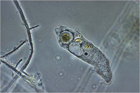
Weds June 18, 2008 at 7:00pm, New Roads School (map) At this meeting, Ed Tarvyd will speak about the diversity of Pacific Marine mammals and their habitats, with an emphasis on species local to Southern California. He will bring and display a variety of examples to illustrate this diversity, including preserved otters, teeth, bones (including some large wale vertebra). Following Ed's presentation, there will be a short program on the photographing diatoms. For many microscopists, diatoms are a favorite subject; often when testing our microscope or new camera system we will use diatoms as our subject. This Wednesday we will learn some new methods for acquiring images through the microscope. |
| Weds July 16, 2008 at 7:00pm, New Roads School (map) At this meeting, Helen Griffin, a forensic scientist and long time associate of MSSC member Ed Jones will give a talk entitled, "The Forensic Examination of Glass." She will be addressing the microscopic identification and characteristics of glass fragments. Helen Griffin has worked as a forensic scientist in the field of Trace evidence examination for 22 years. She authored the chapter Glass Cuts in Forensic Analysis On The Cutting Edge New Methods for Trace Evidence Analysis edited by Robert D. Blackledge, Wiley, 2007. She was a speaker for the FBI/NIJ 2007 Trace Evidence Symposium in Clearwater, Florida where she presented a talk on glass cuts in clothing. Glass is often broken during the commission of a crime. When glass breaks, it flies in all directions, depositing onto anyone in close proximity. This results in small fragments of glass being a fairly common form of forensic evidence. This discussion will briefly cover how glass is collected from a suspect, how it is identified as being glass, and how it is compared to a control source of glass. The downside of glass evidence is that all of the physical and chemical characteristics of glass are class characteristics that belong to a number of different glass sources. Also, broken glass is common in our environment. I will discuss how finding cuts formed by broken glass can add to the strength of finding glass on the suspect's clothing. |
Weds August 20, 2008 at 7:00pm, New Roads School (map) At this meeting, John Koivula will give a talk entitled " The Micro-world of Gems." John was a member of our Society back when we met at the George Page Museum. Currently, he is the senior scientist at the Gemological Institute of America. John is the worlds leading expert on the identification and interpretation of Gem inclusions and is currently working on the third publication of his Atlas of Gem-inclusions. He also produces great photomicrography. In his talk, John will describe and illustrate the importance and significant of inclusions - tiny clues that can be found within the body of a precious stone. We will discover how inclusions are formed and what they mean as to the history and value of a rare gem. For a preview of John's work, visit his website: http://www.microworldofgems.com. John has received numerous awards and honors for his images, including the "Nikon Small World Photomicrography" competition and competitions sponsored by Polaroid and Kodak. His photomicrographs have also graced the covers of Gems & Gemology, Journal of Gemmology, Canadian Gemmologist and Australian Gemmologist, as well as numerous other trade publications. Early in his career, John Koivula realized that making photographs through the microscope was an indispensable tool in his work as a gemologist. Having mastered the intricacies of microscopy and photomicrography, as well as inventing many new photographic techniques over the past thirty years, today John Koivula is the leading authority and foremost photomicrographer of inclusions in gemstones. His photomicrographs not only are visually eloquent and succinct records of what he observes in the microscope, but also are documentary proof for his interpretation of what he observes. |
Weds September 17, 2008 at 7:00pm, New Roads School (map) At this meeting, Simon Malcomber will give a talk entitled " Developmental genetic basis of inflorescence diversity in grasses and immediate relatives (graminoid Poales)." Professor Malcomber is an Assistant Professor with the Department of Biological Sciences at California State University in Long Beach. Prof. Malcomber will take you into the microscopic world of plant development and gene function. Model organisms such as Arabidopsis, maize, and rice provide testable hypotheses of how genes have regulated the evolution of morphological forms within plants. We can test these hypotheses by combining developmental, gene expression and molecular evolutionary analyses of diverse plant species within an explicit phylogenetic framework. The trehalose phophate phosphatase (TPP) gene RAMOSA3 is an upstream regulator or RAMOSA1 in maize and plays an important role in regulating inflorescence branching. Phylogenetic analyses indicate that the gene duplication event producing RAMOSA3 (RA3) and it's paralog SISTER OF RAMOSA3 (SRA) occurred prior to the major radiation of the grass family. In situ hybridization expression analyses in rice and sorghum coupled with phylogenetic reconstructions reveal a complex pattern of expression evolution within the RA3 gene family and point to changes in gene expression and potentially gene function since maize and sorghum last shared a common ancestor. Based on functional analyses in rice, the SEPALLATA gene LEAFY HULL STERILE1 (LHS1) has been linked to both the origin of lemmas within grass flowers and the diversification of the grass spikelet. Phylogenetic analyses indicate that LHS1 and its paralog Oryza sativa MADS5 (OsMADS5) resulted from a gene duplication event at the base of the grass family approximately 80 MYA. LHS1 expression has been detected in lemmas of all grasses tested to date supporting it's role in the development of this structure, but LHS1 expression patterns in grasses are not conserved pointing to additional developmental roles during spikelet development. Analyses in Joinvillea (a close relative of grasses) reveal expression of the LHS1/OsMADS5 precursor in both tepal whorls and in the stamen filament, pointing to potential partitioning of gene function in grasses following the LHS1/OsMADS5 duplication event. Together these analyses show the utility of testing hypotheses derived from model species in non-model species and offer a framework to identify strategies of improving crop yield in diverse species. |
Weds October 15, 2008 at 7:00pm, New Roads School (map) This meeting will start with a slideshow by MSSC member Stuart Warter, entitled, Natural History Adventures in Close-up, Macro- and Telephotography: Part II featuring the unseen or seldom seen lives and loves of birds, bugs, and other assorted critters from the backyard and elsewhere. The second presentation will be by Ken Gregory. Using a Stereo Master projector Ken Gregory will present a "3D dissection of the human heart and thoracic cavity." Come and experience another 3D slide show with Dr. Gregory and his stereo reels. Beautiful views of the dissected heart from outside to inside with commentary on anatomy and function. 3D glasses will be provided. |
Weds November 19, 2008 at 7:00pm, New Roads School (map) This is the annual Exhibition Meeting of the Society. This is one of the best events of the year and is a great deal of fun. Each member is encouraged to bring along an exhibit to share. Anything associated with microscopic subjects is welcome. Your exhibit could be simple, for example you could set up your microscope with your favorite slide. A projector will be provided for those bringing 35mm slides. Posters and display boards are also encouraged, along with the usual sales table. Please remember to bring a label or piece of paper with a brief description of your exhibit. |
No December meeting, instead the MSSC Holiday Banquet, December 14, 2009 at 5:00 pm, Earth, Wind and Flour in Santa Monica. This year, to better satisfy the palates of our members we will be ordering off the main menu. Prices range from $10.00 to $16.00 depending on what you order. Each member must bring cash as there will be no individual checks. Plan on including a 20% gratuity. RSVP to Jim Solliday (see contacts) no later than December 1, 2008. |
WHAT'S
NEW? / MSSC HOME PAGE / MSSC
HISTORY / PROGRAM SCHEDULE /
ITEMS FOR SALE / NEWS
AND EVENTS / ARTICLES & RESOURCES / CONTACT US / HOW TO JOIN / LINKS / MEMBERS
AREA
Cartoons by Nirvan Mullick
Site created and maintained by Leonie
Fedel
Please email comments
© MSSC
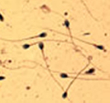 At this meeting, Dr. Edwin Jones Jr, Ventura County Sheriff's Department Forensic Sciences Laboratory and MSSC member will give a presentation entitled, "Sperm Counting and Its Use in Evaluating Postcoital Interval." In fertility studies, sperm counting was used to assess the sperm density of semen. The exposure to semen in the female reproductive tract was evaluated by counting sperm. In the Microscopy of Rape Evidence classroom, sperm counting was used as a quality assurance tool to test the students' ability to find and accurately identify sperm. Sperm counting was also used to evaluate the efficiency of various extraction techniques for sperm from different substrates. Counting sperm on a stained slide with no coverslip or mounting media consistently showed about 20% less sperm than counting the same sample with mounting media and coverslip. Counting sperm on the same slide with 400 and 600 times magnification showed consistently more sperm at 600X. Sperm counting was found to be of assistance in evaluating the postcoital interval (PCI) also known as time since intercourse (TSI) of samples from rape-murder cases. The techniques of sperm counting will be presented so that you can use the data with the published literature in the area of PCI. The extensive literature in the field of PCI will be discussed in relation to the types of sampling methods (swabs, smears on microscope slides and vaginal washings). One paper from this literature stands out as being useful for assessing PCI because the female volunteers were active after sex and it used 12 or more sperm to define the 4+ rating of sperm (1). The author will present a method for obtaining better data from the samples normally encountered in sexual assault investigations. This method involves counting the number of sperm and the number of nucleated squamous epithelial cells in each field of view.
 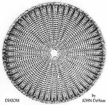 Dr. Jan Hinsch, former director of the Microscopy lab at E.Leitz Rockleigh (Wild Leitz, Leica) will give a presentation at this meeting entitled, "About Microscopes and Diatoms." Dr. Hinsch's work in the field includes a career as the Director of the Laboratory for Applied Microscopy at Leica Microsystems, and service as an instructor at the prestigious Marine Biological Laboratory course in Analytical and Quantitative Microscopy. Hinsch was the recipient of the Ernst Abbe Memorial Award in 2002 recognizing his work as a leader in light microscopy technology and education. The following is an abstract provided by Jan that will be presented at our March meeting. "Diatoms fascinate microscopists. Almost everyone is attracted to the formal beauty of these microscopic plants. Moreover, to the person with an interest in the physics of the microscope the silica shells of diatoms with their repetitive, ordered fine structure are suitable objects for various kinds of enlightening diffraction experiments. In places where microscopy is taught comprehensively, the study of diatom structure is part of the curriculum intended to deepen the understanding of terms like resolution and corrections for chromatic and spherical aberrations of microscope objectives. Often progress in microscope optics and illumination methods, such as DIC, has been demonstrated first on diatoms. I like to reference some of these techniques in my talk with emphasis on Reflection Interference Contrast (RIC). To the diatomist this is a relatively new technique that, under favorable circumstances, enhances contrast and thereby permits to achieve resolution close to the theoretical limits imposed by the lens aperture and the wavelength of light. RIC images are also more representative of the surface of diatoms than are transmitted light images and in this respect resemble SEM micrographs. The availability of interesting software, much of it in the public domain, is a major reason why I replaced my trusted 35mm microscope camera with a digital model. Between Photoshop Elements, ImageJ and CombineZ I can acquire, enhance and analyze my images with ease."